The aim of the curriculum is to provide a correlated sequence of systematic instruction and clinical experience in the Science of Radiologic Technology. The program is based on two years of study with a maximum of forty (40) hours of combined clinical and academic instruction per week.

For a student to progress through the program and graduate he / she must maintain a 75% average or above in each individual course as well as an overall grade. Evaluation will consist of, written and practical examinations and observation.

Program Outline - Year 1

Trimester # 1

  • RT 101 – Intro to Radiologic Technology
  • RT 102 – Medical Terminology 1
  • RT 103 – Patient Care and Pharmacology 1
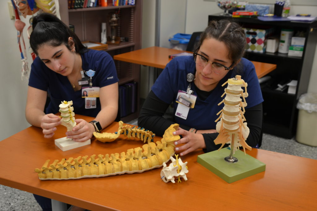
  • RT 104 – Anatomy & Physiology 1
  • RT 105 – Procedures 1
  • RT 106 – Procedures Lab 1
  • RT 107 – Physics & Equipment 1
  • RT 109 – Image Critique 1
  • RT 112 – Radiation Protection and Biology 1
  • RT 110 -Clinical Experience 1

Trimester # 2

  • RT 203 – Patient Care and Pharmacology 2
  • RT 204 – Anatomy & Physiology 2
  • RT 205 – Procedures 2
  • RT 206 – Procedures Lab 2
  • RT 207 – Physics & Equipment 2
  • RT 208 – Exposure & Processing 1
  • RT 209 – Image Critique 1 continued
  • RT 210 – Clinical Experience 2
  • RT 212 – Radiation Protection & Biology 2

Trimester # 3

  • RT 310 – Clinical 3

Program Outline - Year 2

Trimester # 4

  • RT 408 – Exposure & Processing 2
  • RT 404 – Anatomy & Physiology 3
  • RT 405 – Procedures 3
  • RT 406 – Procedures Lab 3
  • RT 411 – Pathology
  • RT 410 – Clinical Experience 4

Trimester # 5

  • RT 512 – Senior Review
  • RT 510 – Clinical Experience 5
  • RT 409 – Image Critique 2

Trimester # 6

  • RT 610 – Clinical Experience 6

Trimesters run tentatively

1 = 3rd Week in August – 3rd Week in December
2 = 1st Week in January – 4th Week in May
3 = 1st Week in June – 2nd Week in September
4 = 3rd Week in September – 3rd Week in December
5 = 1st Week in January – 4th Week in April
6 = 1st Week in May – 2nd Week in June

Course Descriptions - Year 1

The students are given an overview of the history of radiology. The policies of the radiology program are reviewed and discussed. An understanding of the healthcare team is established. The students are made aware of the organizational structure of the radiology department and the hospital. The students are taught about accreditation, certification, licensing, professional organizations, and career advancements in radiology. The students are introduced to medical ethics and medico-legal principles.

The art of effective communication and body language is discussed. The importance of medical asepsis in the radiology department is realized. The students learn how to admit a patient to the radiology department and how to properly move and transfer patients. Vital signs and oxygen administration are learned and practiced. The students are taught to care for patients with various problems (e.g. skull or spinal injuries, pediatric or geriatric patients, shock, etc.). Proper methods of the administration of enemas and caring for patients with various types of tubes are discussed. Surgical aseptic technique, proper skin preparation, and venipuncture is learned. The student is taught how to assist with drug and contrast media administration. Various contrast complications and anaphylactic reactions are discussed. Transmission of microorganisms and isolation techniques are learned.

The students will learn the origins, word building, terminology related to the various systems of the body and medical abbreviations and symbols that are routinely used in radiography.

The students are introduced to the anatomical terms and units of body structure. An understanding of the functions of cells and their components is established. Students are introduced to the various classifications of bones, joints, and muscles. They must learn all the individual bones of the extremities, spine, and thorax.
To provide the students with the knowledge of the structure and function of the human body with emphasis on radiographic aspects of the cranium, facial bones, sinuses, and mandible and the urinary, digestive, respiratory and the integumentary systems.
The students are introduced to anatomical and positioning terminology and abbreviations that are routinely used in radiology. They are taught how to perform basic positioning skills for radiography of the chest, upper and lower extremities, shoulder girdle, pelvic girdle, spine, and bony thorax.
The students are taught the proper patient preparation, various contrast studies and positioning skills for abdomens, upper GIs, large bowel, gallbladder, and IVP examinations. Portable radiography, long bone measurement, arthrography, foreign body localization, trauma examinations and pediatric examinations.
The students are taught how to perform radiographic examinations using laboratory simulations prior to performing actual exams on patients.
The students are introduced to the concepts of radiation, fundamentals of mathematics, and units of measurement. An understanding of the structure of matter and the basic components of the atom is taught. Students will learn about the principles of electromagnetic radiation, electricity, and magnetism. An understanding of basic circuits, generators, motors, transformers, and rectifiers is established. The students will comprehend the components of the x-ray tube, the basic circuitry, and the production of x-radiation.
To provide the students with the knowledge of various imaging systems and their applications to radiography. The students will obtain an understanding of the general principles of equipment that uses ionizing radiation and equipment that uses non-ionizing radiation. X-ray production and emission spectra will be discussed. The five interactions with matter will be reviewed. Image intensification and fluoroscopic image monitors will be learned. Tomographic and mammmographic equipment will be analyzed. The students will be introduced to computer science in radiology and the use of computers for digital imaging and computed radiography. MRI and ultrasound will also be discussed.
The students are first introduced to basic radiation protection practices. Five possible interactions of radiation with matter are learned. Sources of radiation, radiation units, and the cardinal principles of protection are discussed. Cell biology and possible radiation effects are established. The early and late somatic effects of radiation are learned. The design of x-ray facilities is discussed and means of reducing exposure to patients and personnel are understood. Radiation monitoring, detection, and measurement is also learned.
The students will first be introduced to the production of x-rays and general radiographic terms. Time will be spent on basic mathematic principles and applications to radiology. The characteristics of a radiographic image and the factors that affect them will be discussed. Image unsharpness, distortion, milliamperage, timing devices, source-image receptor distance, kVp, tissue interactions, beam restriction, and radiographic grids will all be discussed at length.
The students will learn the importance of correct patient identification, dates, and markers on all radiographs. Proper positioning, exposure factors, source-image receptor distance, collimation, shielding, structures shown for all the various examinations of the chest, upper and lower extremities, shoulder girdle, pelvic girdle, spine, and bony thorax will be discussed in detail.
The clinical experience is where the student has the opportunity to apply what has been learned in the Radiographic Procedures classes. Students will be expected to perform exams on patients after they have been covered in the didactic portion of the training. The student will use a step method where they will practice, perform preliminary competencies, formal competencies and a capstone of a Terminal comp at the end of each clinical experience.

Course Descriptions - Year 2

In this section of the course the student will have a basic understanding of the endocrine, reproductive, circulatory, lymphatic, nervous and sensory systems (organs).
The student will be taught the proper positioning for all head work exams. Areas covered are facial bones, nasal bones, paranasal sinuses as well as orbits, mandible and other special views. Surgical procedures in which mobile fluoro, C-arm, is used along with the retrograde room and all other operating room procedures. Special procedures for the nervous system and circulatory systems and specialized modalities are discussed.
The students are taught how to perform radiographic examinations using laboratory simulations prior to performing actual exams on patients.
To enable the students to recognize the difference between good diagnostic radiographs and poor quality radiographs. The students will explain how to improve the diagnostic quality of various radiographs while viewing them in class. The student will learn the importance of correct patient identification, dates, and markers on all radiographs. Proper positioning, exposure factors, source-image receptor distance, collimation, and shielding for all the various examinations of the abdomen, digestive system, urinary system, skull, portable radiography, pediatric radiography, mammography, and special procedures will be discussed in detail.

To acquaint the students with basic medical terminology used to describe various pathologic conditions occurring in the human body and introduce the students to some specialized imaging techniques. The students will be able to classify more common diseases in terms of their penetrability by x-rays and become familiar with the changes in technical factors required for obtaining optimal quality radiographs on patients with various underlying pathologic conditions.

The students will learn various pathologic conditions of the respiratory system, skeletal system, gastrointestinal system, urinary system, reproductive system, nervous system, endocrine system, hemopoietic system, and cardiovasular system.

To provide the students with the knowledge of the factors that govern and influence the production of the radiographic image on the film, processing, and QA/QC that Is used in radiology. The students will understand the meaning of good radiographic quality and how each of the exposure factors effect it. The construction of x-ray film, proper storage and handling, and sensitometry will be learned. Designing technique charts will be discussed. Multiple technique problems will be introduced and solutions will be established. Processing and artifacts will be discussed in detail. QA and QC will be defined. Testing and implementing a program will be taught.
This is a capstone course that will review major point that was covered during the student’s didactic & clinical education. The focus of this course will to resume preparation, interview skills and mock Registry exams. The intent of this course is to prepare the student for the ARRT exam and for employment after graduation.
The clinical experience is where the student has the opportunity to apply what has been learned in the Radiographic Procedures classes. Students will be expected to perform exams on patients after they have been covered in the didactic portion of the training. The student will use a step method where they will practice, perform preliminary competencies, formal competencies and a capstone of a Terminal comp at the end of each clinical experience.

Graduation Requirements

A student who successfully completes the following requirements of the school will (1) qualify for graduation and (2) be permitted to sit for the American Registry of Radiologic Technologist Examination for national certification and state licensing.

  1. Complete all academic courses and the clinical competency program according to the established criteria. A student with incomplete records in either of these areas shall not be granted a certificate and will not be authorized by the Educational Director as meeting the educational requirements for certification by the American Registry of Radiologic Technologists.
  2. Complete two (2) years of training in Medical Radiography with a minimum average of “75%” in all subjects. Failure on the student’s part to fully complete the program prior to the graduation date will require the student to remain after graduation for completion.
  3. Demonstrate competence in all routine procedures and techniques.
  4. Meet all financial obligations to the school.
  5. Be of good moral character and able to communicate effectively with patients and personnel in an ethical manner.
  6. Be an asset to the profession in accordance with faculty and administration evaluations.